Musculoskeletal System for the MCAT: Everything You Need to Know
/Learn key MCAT concepts about the musculoskeletal system, plus practice questions and answers
(Note: This guide is part of our MCAT Biology series.)
Table of Contents
Part 1: Introduction
Part 2: Types of muscle
Part 3: Muscle contraction
Part 4: Additional connective elements
a) Bone structure
b) Bone maintenance
c) Tendons, ligaments, and cartilage
Part 5: Skin
a) Structure
b) Homeostasis
Part 6: High-yield terms
Part 7: Passage-based questions and answers
Part 8: Standalone questions and answers
----
Part 1: Introduction
The musculoskeletal system supports the body and allows us to move. Therefore, it is important to understand its structure as well as how muscle contraction typically occurs. In this guide, we will provide you with the content you need to know for the MCAT. There are many terms associated with the musculoskeletal system, so a foundational understanding is crucial for retaining the content. At the end of this guide, there is an MCAT-style musculoskeletal practice passage and standalone questions to test your knowledge and show you how the AAMC likes to ask questions.
Let’s get started!
----
Part 2: Types of muscle
There are many different types of muscle throughout the body, and all types have various forms and functions. In this section, we will introduce you to three different types of muscle: skeletal, cardiac, and smooth muscle.
Skeletal muscle is the tissue responsible for voluntary movement. It is consciously controlled and innervated by the somatic nervous system innervations (more to follow in part three). Skeletal muscles are striated, or striped, and are multinucleated.
There are two types of skeletal muscle fibers: slow-twitch fibers and fast-twitch fibers. These two forms of muscle differ in their contractile velocity, or how rapidly they are capable of contracting to produce movement. Slow-twitch fibers are sometimes referred to as red fibers or type I fibers and have low contractile velocity. They are red because they carry lots of myoglobin, an oxygen carrier that only has one subunit of hemoglobin. Slow-twitch fibers also have large amounts of mitochondria and are slower to fatigue. Fast-twitch fibers are also called white fibers or type II fibers and have high contractile velocity. They contain much less myoglobin and are therefore lighter in color. They contract rapidly but tire quickly.
Skeletal muscles are additionally prone to suffering fatigue. Fatigue is a consequence of suffering oxygen debt: a disparity between how much oxygen the muscles require to produce sufficient energy (in the form of ATP), and how much oxygen is being provided through breathing. (For more information on the role of oxygen in energy production, be sure to refer to our guide on carbohydrate metabolism.)
The contraction and relaxation of skeletal muscle around the body perform an additional function in moving fluids. Blood and lymph, residing in blood vessels and lymph vessels, may be periodically “squeezed” due to the motion of the surrounding skeletal muscles. This movement assists in returning fluid from the periphery of the body, where blood pressure tends to be low.
A sarcomere is a unit of skeletal muscle. Sarcomeres are composed of two different strands of protein filaments colloquially called thick and thin filaments. These filaments interweave with each other in what is referred to as the contractile apparatus. Thick filaments are composed of myosin, while thin filaments are made of actin. Troponin and tropomyosin attach to the actin, which works together with myosin to contract muscle. Sarcomeres are connected end-to-end to form myofibrils. Each myofibril is surrounded by sarcoplasmic reticulum, a specialized membranous organelle that contains high concentrations of Ca²⁺ ions. These Ca²⁺ ions play an important role in muscle contraction.
Smooth muscle is involuntary and is controlled and innervated by the autonomic nervous system. Smooth muscle lines the digestive tract, bladder, uterus, blood vessel walls, and other regions of the body responsible for transportation of material or peristalsis. Both skeletal and smooth muscles are made of thick and thin filaments. Both skeletal and smooth muscles respond to input from the nervous system to undergo contraction. However, in smooth muscle, the myosin and actin fibers are not organized into sarcomeres. Additionally, smooth muscle can exhibit myogenic activity, which is contraction without nervous system input, and has led to the common belief that there is a “second brain” in your gut! In addition, smooth muscles are not striated and only have a single nucleus at the cell center.
Cardiac muscle is unique to the heart and contains characteristics of both skeletal and smooth muscle. Like skeletal muscle, cardiac muscle is striated and composed of sarcomeres. Like smooth muscle, cardiac muscle is involuntary, and each muscle cell only contains one nucleus. Cardiac muscle cells are separated by intercalated discs. These intercalated discs contain many gap junctions that allow for ions to rapidly flow and action potentials to be quickly propagated throughout the heart, resulting in efficient and coordinated contraction. While action potentials in neurons require the propagation of signals through one nerve cell at a time, the unhindered flow of calcium ions through gap junctions allows for synchronized contraction.
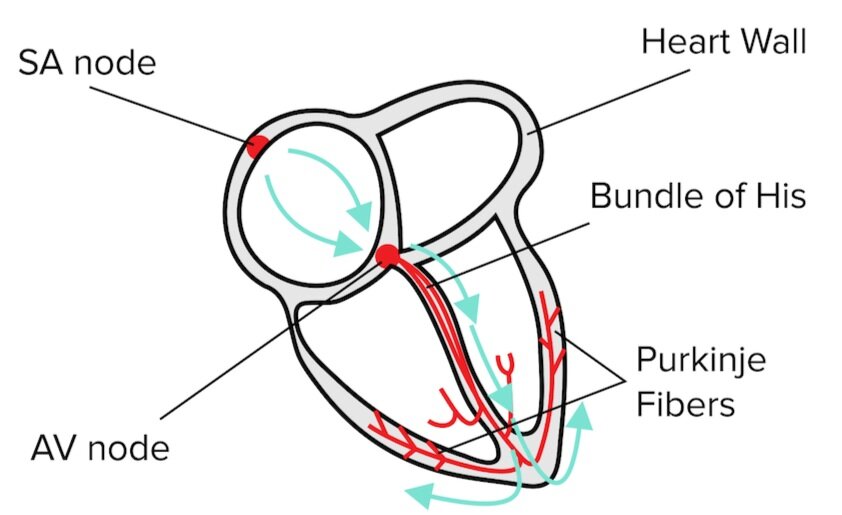
Cardiac muscle cells exhibit myogenic activity, or electrical activity independent of the brain that regulates the rhythm of the heart. These electrical signals start at a cluster of specialized electrical cells at the top of the heart, known as the sinoatrial node. The electrical signals then propagate throughout the heart, triggering muscle contraction. From the sinoatrial (SA) node, the signal travels through the atrioventricular (AV) node and the Bundle of His. Lastly, the signal spreads through the Purkinje fibers: branching fibers in the walls of the heart ventricles that induce contraction in the cardiac muscle.
Figure: A depiction of the flow of electricity through the heart.
| Skeletal | Smooth | Cardiac |
|---|---|---|
Gain instant access to the most digestible and comprehensive MCAT content resources available. 60+ guides covering every content area. Subscribe today to lock in the current investments, which will be increasing in the future for new subscribers.


