Digestive and Excretory Systems for the MCAT: Everything You Need to Know
/Learn key MCAT concepts about the digestive and excretory systems, plus practice questions and answers
(Note: This guide is part of our MCAT Biology series.)
Table of Contents
Part 1: Introduction to the digestive and excretory systems
Part 2: Digestive system
a) Gastrointestinal anatomy
b) Accessory organs
c) Enzymatic digestion and absorption
Part 3: Excretory system
a) Urine production
b) The nephron
c) Endocrine control
Part 4: High-yield terms
Part 5: Passage-based questions and answers
Part 6: Standalone questions and answers
-----
Part 1: Introduction to the digestive and excretory systems
The body requires energy to function and maintain homeostasis. How does our body absorb the nutrients it needs and excrete the waste it produces?
The digestive and excretory systems work together to break down the food we eat into absorbable nutrients and expel waste. A significant amount of biology questions on the MCAT directly cover content described in this guide—including the structure and function of the kidney. Having a thorough understanding of these systems is a definite advantage now and in your future medical career.
Throughout this guide, you will find several bolded terms that are important to understand and recall. At the end of this guide, you will also find several MCAT-style questions for you to test your knowledge.
Let’s jump right in!
-----
Part 2: Digestive system
a) Gastrointestinal anatomy
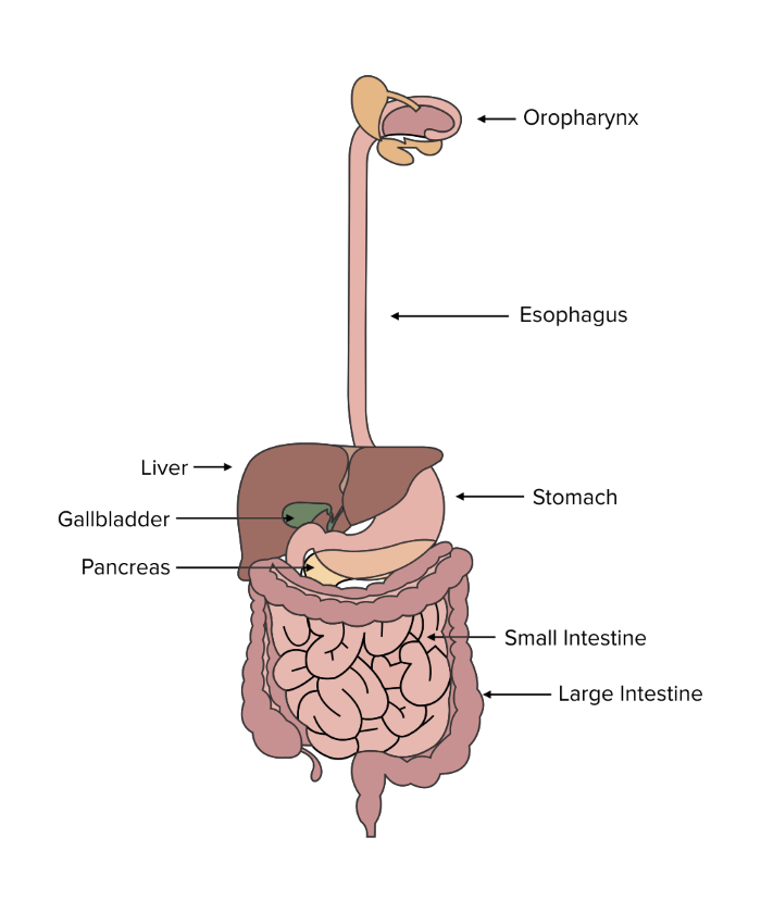
The digestive tract is a long, tube-like structure that runs through the entire torso and abdomen of the human body. Food that enters the digestive tract at the mouth undergoes a series of digestive steps as useful nutrients are absorbed into the bloodstream, and any unabsorbed nutrients exit as waste.
Believe it or not, digestion starts at the mouth. The mouth is responsible for both the mechanical and chemical digestion of food. Mastication, or chewing, is the mouth’s primary method of mechanical digestion. Mechanical digestion results in the breaking down of large particles of food into smaller pieces, increasing the total surface area. The increased surface area aids in chemical digestion performed by salivary enzymes. The salivary glands of the mouth produce enzymes, known as salivary amylase and salivary lipase, which begin to break down the chemical bonds of sugars and lipids in the food. As food doesn’t stay for very long in the mouth, the degree of digestion is quite limited but will continue further along the digestive tract.
Figure: Anatomy of the digestive tract.
In preparation for swallowing, the tongue rolls food into a ball-shaped mass known as a bolus. The bolus is then moved into the oropharynx, where muscles work to push the bolus out of the mouth.
During swallowing, food passes through the pharynx, a tube-shaped structure that is shared between the respiratory and digestive systems. To prevent food from entering the lungs, a flap-like structure known as the epiglottis automatically closes the opening of the larynx. As a result, food can safely travel down the pharynx and into the esophagus.
The esophagus connects the mouth to the stomach and has two sphincters: the upper and lower esophageal sphincters. (Recall that sphincters are simply ring-shaped muscles that can open and close by contracting.) These sphincters regulate the entrance of food into the esophagus and the stomach, respectively.
Perhaps surprisingly, humans lack voluntary control of the esophagus. Only the upper third of the esophagus is composed of skeletal muscle, which allows us to swallow our food. Our food moves down the rest of the esophagus through wavelike movement created by the smooth muscle occupying the rest of the tube. This movement is known as peristalsis. The movement is assisted by the presence of saliva that has been mixed with the bolus, which serves an additional function as a lubricant.
As our food moves down the esophagus, it eventually reaches the stomach. The stomach is where much of the chemical digestion of food occurs. Several glands in the stomach lining secrete acid chemicals and other enzymes to initiate chemical digestion.
Hydrogen ions are secreted by parietal cells within the stomach.
Pepsinogen is secreted by chief cells within the stomach. Pepsinogen is a zymogen, an inactive form of the enzyme pepsin that has to be cleaved with hydrogen ions to become active pepsin.
Pepsin, the activated form of pepsinogen, is responsible for breaking proteins down into amino acids.
Gastrin is secreted by G-cells located within pyloric glands. Gastrin stimulates the secretion of hydrochloric acid (HCl). The stomach contains a large amount of hydrochloric acid, producing an acidic environment.
Mucus is secreted by goblet cells. This mucus protects the lining of the stomach and prevents the highly acidic environment from damaging the stomach itself.
Chief cells and parietal cells are found together in gastric glands. Assisted by the mechanical churning of muscles surrounding the stomach, the enzymes and acid secreted in the stomach digest food into chyme. Chyme is a thick, heterogeneous mixture of food and digestive enzymes that undergoes further digestion and absorption in the small intestine.
The resulting chyme travels to the small intestine. The small intestine is responsible for the digestion and absorption of nutrients. The small intestine consists of three different sequential sections: the duodenum, jejunum, and ileum. Alkaline juices produced by the pancreas, an accessory organ, are secreted into the duodenum and neutralize the highly acidic contents arriving from the stomach. Chyme initially travels through the duodenum, where the presence of chyme in the duodenum initiates the release of many enzymes from several accessory organs that are discussed later in this guide. These enzymes perform the bulk of chemical digestion as the food passes through the digestive tract.
After passing through the duodenum, the chyme makes its way to the jejunum and ileum for absorption. Characteristic of these regions are microvilli, which are small, finger-like structures that increase the surface area of the intestine to promote absorption. Here, the nutrients that are present in the digested mixture are absorbed into epithelial cells and transported into the capillaries for use. Most small molecules that aren’t hydrophobic, such as water, simple sugars, most vitamins, and amino acids, are simply absorbed through facilitated diffusion and secondary active transport into epithelial cells and then into capillaries.
For molecules that are hydrophobic, including the fat-soluble vitamins K, A, D, and E, special structures called lacteals exist to allow these molecules into the lymphatic system, which eventually empties into the left subclavian vein. For more information on the absorption and digestion of fats, be sure to refer to our guide on lipid and amino acid metabolism.
Finally, after passing through the small intestine, the mixture makes its way to the large intestine. The majority of nutrients—such as amino acids, carbohydrates, and vitamins—have been absorbed in the small intestine.
The large intestine is responsible for absorbing water and leftover salts. It also contains gut flora, colonies of beneficial bacteria that reside within the body and assist in the production and absorption of nutrients such as vitamin K. After passing through the large intestine, the only material that remains after absorption is waste, which is consolidated into fecal matter.
Fecal matter is stored in the last segment of the large intestine. When the body is ready to eliminate waste, it is pushed through the rectum and exits the body through the anal sphincters.
The movement and peristalsis of material through the digestive tract is largely governed by the enteric nervous system: a subdivision of the autonomic nervous system. The nerve endings within the enteric nervous system enervate sections of the smooth muscle lining the digestive tract, thus governing the rate of digestion as food passes through the body.
b) Accessory organs
Although these accessory organs do not form any portion of the tubelike digestive tract, the liver, gallbladder, and pancreas are important accessory organs that aid in chemical digestion.
Located in the upper-right quadrant of the abdomen, the liver plays many important roles in aiding digestion. One of the most important functions the liver serves is producing bile. Bile is a mixture composed of different salts, cholesterol, and bilirubin pigment.
Gain instant access to the most digestible and comprehensive MCAT content resources available. 60+ guides covering every content area. Subscribe today to lock in the current investments, which will be increasing in the future for new subscribers.


