Cells for the DAT
/Learn key DAT concepts related to cells, plus practice questions and answers

Everything you need to know about cells for the dat
Table of Contents
Part 1: Introduction to cells
Part 2: Cell theory
Part 3: Cell structure and organelles
a) Organelles
b) Cytoskeleton
c) Higher structure in multicellular organisms
Part 4: Cell membrane and components
a) Phospholipid bilayers
b) Fluid mosaic model
c) Major components of the cell membrane
d) Transporters
Part 5: Cell division
a) Mitosis
b) Meiosis
c) Dysfunctional cell growth
Part 6: High-yield terms
Part 7: Questions and answers
----
Part 1: Introduction to cells
Cells serve as the fundamental units of living organisms and hold significant importance in the context of the biology section of the DAT. Cells can be directly examined, and they form the core of various biological concepts discussed in passages and experiments. Therefore, establishing a solid understanding of cellular biology is crucial. Key terms have been bolded throughout this guide, accompanied by definitions at the end. Additionally, practice questions and answers are included to test your learning.
----
Part 2: Cell theory
In 1655, the English scientist Robert Hooke conducted a microscopic examination of cork, leading to the formulation of the three fundamental principles of cell theory:
All living organisms consist of one or more cells.
Cells represent the fundamental unit of life.
New cells emerge through the division of pre-existing, living cells.
Over the years, cell theory has evolved to incorporate new discoveries, notably acknowledging DNA as the genetic information within each cell and recognizing the transmission of DNA from one cell to another.
There are two main cell types that serve as the basis for classifying all living organisms: prokaryotes and eukaryotes. Viruses, however, fall outside the realm of living entities. For further details on viruses, see our guide on microbes.

FIGURE 1: PROKARYOTE, EUKARYOTE, AND VIRUS CELLS
The DAT commonly tests the differences between prokaryotes and eukaryotes. As such, you should know the details listed in the table below.
| Prokaryotes | Eukaryotes |
|---|---|
----
Part 3: Cell structure and organelles
a) Organelles
Cells consist of organelles that help them function. Organelles are structures inside of a cell that perform specific functions. The image below depicts these organelles in a eukaryotic cell.
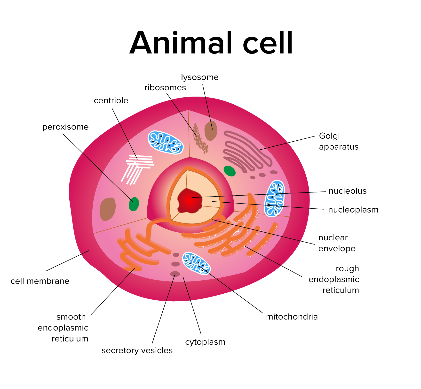
FIGURE 2: LABELED ANIMAL (EUKARYOTIC) CELL
Key organelles to remember from the diagram above include the nucleus, mitochondria, ribosomes, endoplasmic reticulum, lysosomes, Golgi apparatus, peroxisomes, and vacuole. You should know the function of each of these organelles and how they work together in the cell.
| Organelle | Function |
|---|---|
The nucleus is a membrane-bound organelle that controls the activities of the cell. DNA is housed in the nucleus. Inside the nucleus, there is a spherical structure called the nucleolus. The nucleolus is the site of ribosome synthesis.
Ribosomes are enzymes involved in the production of proteins, and are covered more in-depth in our guide on DNA and RNA. Ribosomes can be found in both the cytoplasm and the endoplasmic reticulum. The cytoplasm is a gel-like substance found inside the cell, within which the organelles are contained. The endoplasmic reticulum (ER) is an organelle that is involved with the synthesis of proteins and lipids. It consists of a smooth and rough section, with lipids being synthesized in the smooth ER and proteins being made in the ribosomes of the rough ER. The Golgi apparatus then packages proteins and distributes them throughout the cell.
Another important organelle is the mitochondria. The mitochondria is double-membraned, and produces ATP to power the cell. The mitochondria is the site of cellular respiration, a topic covered in our guide on metabolism. Lysosomes are membrane-bound organelles that “lyse,” or break down, cellular waste. Peroxisomes are another type of organelle. These organelles are involved in lipid metabolism and help with the detoxification of the cell via hydrolysis. Finally, vacuoles are membrane-bound organelles that store nutrients and other materials. They are also used to transport materials between organelles.
b) Cytoskeleton
Much like your body has a skeleton that supports its structure and facilitates movement, cells have a skeleton that performs the same functions. The cytoskeleton consists of proteins that function to support the cell’s structure, help with movement, and transport various materials throughout the cell.
Three key cytoskeleton structures to know are microtubules, intermediate filaments, and microfilaments. Microtubules are the largest of these structures, and are made of a protein called tubulin. They function to provide structural support to the cell. In addition to providing structural support, microtubules can be arranged into different structures such as centrioles, cilia, and flagella.
| Component | Structure | Function |
|---|---|---|
Centrioles are cylinders of microtubules that play a role in cell division. Cilia and Flagella are hairlike structures found outside the cell, with flagella being longer than cilia. Cilia are only found in eukaryotic cells, and play a role in cell locomotion, as well as the movement of substances and fluids throughout the cell. Flagella can be found in prokaryotic cells or eukaryotic cells, and is involved in cellular locomotion as well. In prokaryotic cells, however, flagella is made of flagellin, not microtubules. One important fact to know about eukaryotic cilia and flagella is that they have a 9+2 structure. This means that they are made up of an outer ring of 9 pairs of microtubules around an inner ring of 2 microtubules.
Intermediate filaments are smaller than microtubules. Similar to microtubules, though, intermediate filaments are important for the structural support of a cell. Microfilaments are smaller than both intermediate filaments and microtubules. Their main function is movement, but not the same as how cilia and flagella move the cell. While cilia and flagella are located outside of the cell, microfilaments are found inside the cell. Microfilaments are made of actin filaments, and they function to contract muscle and mix up the cytoplasm of cells in a process called cyclosis.
c) Higher structure in multicellular organisms
In multicellular organisms such as ourselves, individual cells can specialize to perform specific functions. Cells can associate themselves with other cells to further perform important functions.
These associations are especially important in organ systems. For example, cells are able to first organize themselves into tissues. Multiple tissue types together associate themselves with organs. Finally, multiple organs and organ systems make up the entire organism.
Constituent cells may adopt even more specialized functions based on the function of a specific organ or tissue. The shape of a cell can be a major difference between specialized cells. Epithelial cells, for instance, tend to be cube-shaped, while squamous cells are usually flat and thin. Epithelial cells tend to be found in the linings of body cavities and vessels, while squamous cells are found in areas of the body that can be worn down (such as the outer surface of the skin).
The connections between cells, known as intercellular junctions, can also vary depending on their function. Common intercellular junctions include:
Tight junctions, which form a sealed connection between cells and do not allow fluid to pass between cells. These junctions are commonly found in storage organs such as the kidney and stomach.
Gap junctions, which form passages through which ions and nutrients can flow into neighboring cells. These junctions are found in cardiac muscle, where they allow for the rapid flow of calcium ions during contraction.
Desmosomes, which are formed by anchoring the cytoskeletons of adjacent cells together. These junctions are found in tissues that require tensile strength, such as the skin and muscles.
----

Gain instant access to the most digestible and comprehensive DAT content resources available. Subscribe today to lock in the current investments, which will be increasing in the future for new subscribers.